You may have undergone an MRI in the past to identify the root cause of pain, rule out an injury, or for another diagnostic reason. But if you haven’t been through this procedure, something you may have heard about MRIs is that they take a long time. But why, and what does that mean? Understanding why MRIs take longer than other scans can also show how important they are, especially after something like an injury from a car accident that can have serious consequences.
How MRIs Work
A magnetic resonance imaging (MRI) scan is a common procedure throughout the world because it is non-invasive while providing a lot of information on the body. The scan relied on a strong magnetic field and radio waves in order to create intricate cross-sectional images of the organs and tissues in the body.
Unlike x-rays and CT scans, an MRI does not use radiation, which can damage cells. Instead, it uses magnets and radio waves which do not have the same sort of side effects. The magnets used are so strong they could pick up an entire car if used in a standard way. However, in an MRI, the magnetic field is used to realign protons, turning them to face new directions. When the magnetic field is turned off, they return to their previous position. The radiofrequency picks up this movement and allows a computer to visualize everything from soft tissue, bones, and organs like the heart and brain.
The invention of MRI scans revolutionized medical practices, allowing for examination of the inside of the human body in a way that was not previously possible. This technology can be used for a wide range of diagnostics, but some examples include:
- Brain and spinal cord anomalies
- Tumors, cysts, and other anomalies throughout the body
- Breast cancer screenings
- Injuries to the joints, like the back and knees
- Certain types of heart problems
- Diseases of the liver and other abdominal organs
- Evaluation of pelvic pain in women, especially from fibroids and endometriosis
- Suspected uterine anomalies in women undergoing evaluation for infertility
Preparing for an MRI
There is usually no preparation required for an MRI.
When you arrive at the facility that is performing your MRI, you will be asked to change into a gown and remove any metal, including piercings or glasses. Because of the forceful magnets used during the scan, metal objects cannot enter the scanner. A person will usually be unable to have an MRI performed if they have any metal in their body, including bullets, shrapnel, cochlear implants, aneurysm clips, pacemakers, or screws in the bones.
If you suffer from claustrophobia, it is important to share this with your provider. You may be given medication prior to the MRI to ease your anxiety or sedate you.
Depending on the purpose of your MRI, you may also be given an injection of intravenous contrast liquid to help improve the visibility of certain tissues.
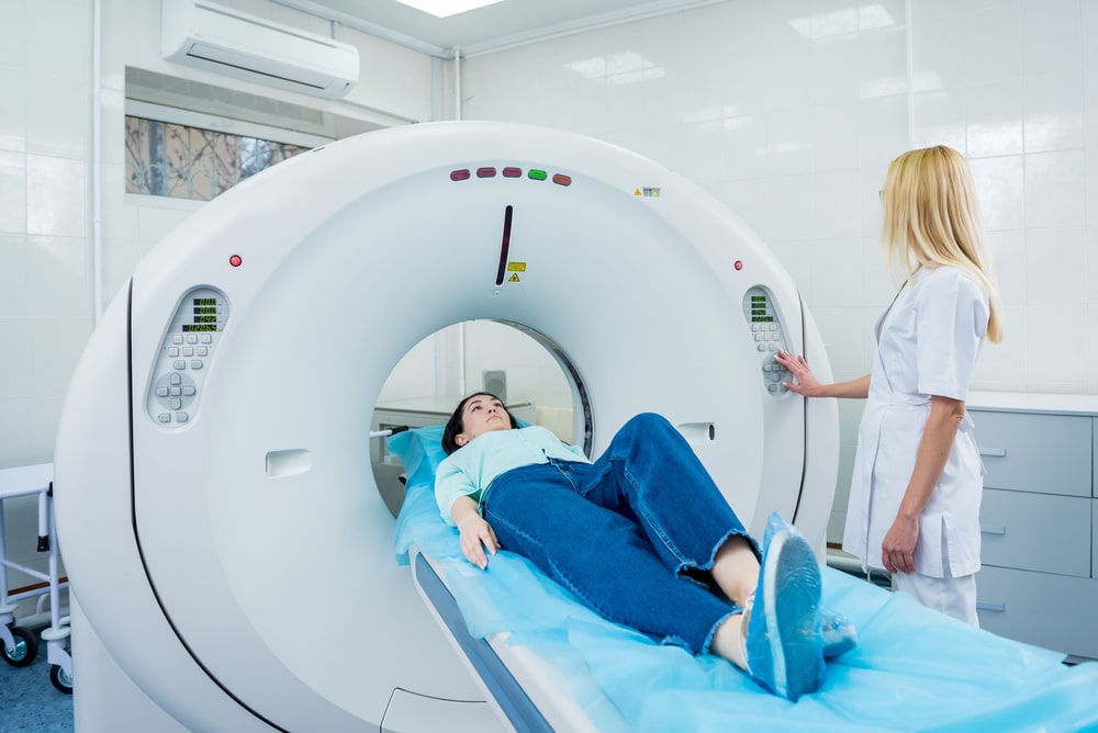
Once you are ready, the radiologist will talk you through the procedure and answer any questions before helping you onto the scanner table. Staff will help you be as comfortable as possible. You will be provided headphones or earplugs to help block out the sound of the scanner and hear instructions during the scan.
Getting an MRI Scan
Once you have entered the scanner, which is a large metal tube, the technician will communicate via an intercom or headphones to ensure you are comfortable and ready to start.
The most difficult part of a scan is usually that you have to stay entirely still. Any movement can disrupt the images, like a camera trying to take a picture of a moving object. The scanner will make loud clanging noises that may sound scary but are totally normal. You may also be asked to hold your breath at certain points.
If you feel uncomfortable at any point, the same intercom can be used to speak to the technician and request the scan be stopped.
After the scan, a radiologist can examine the images to ensure they are of good enough quality. If so, you will be released. Unless you have used a sedative, you should be able to drive home immediately.
Side Effects of an MRI
It is very rare for someone to experience side effects from an MRI. People who suffer from claustrophobia may need time to mentally recover, especially if they did not use medication prior.
The contrast dye can cause some mild side effects like nausea, headaches, and pain or burning at the injection site. It is also possible, but not common, to have an allergy to the contrast that causes hives or itchy eyes.
MRI Length By Type
MRIs are known to take a long time compared to other scans, like x-rays. The exact length of time may vary by the area being scanned along with the type of injury which required different types of MRI scans.
Knee MRIs
An MRI of the knee may be used to identify weakness, swelling, or bleeding around the joint. This will usually take about 30 to 60 minutes. During the scan, small devices containing coils may be placed around the knee to improve image quality.
Shoulder MRIs
An MRI of the shoulder will usually take between 15 and 45 minutes.
Head and Brain MRIs
You may be sent for an MRI of your head to identify any issues affecting your brain. This usually takes about 30 to 60 minutes, depending on if contrast dye is needed. A scan without dye will usually take less time. Some procedures, like a limited brain MRI, could be done in as little as 5 minutes.
Lumbar Spine MRI
Your lumbar spine, or lower back, may need to be scanned to identify causes of back pain that did not appear on other scans, like conditions involving the spinal cord. Without dye, this may take 30 to 60 minutes. With dye, you should expect something closer to 45 to 80 minutes.
Cervical Spine MRIs
An MRI of your upper back and neck, or cervical spine, can take as little as 20 minutes or as much as 45 minutes. It is common to have a plastic coil placed on the top of your neck to help produce a clearer picture.
Cardiac MRIs
For an MRI of your heart, the scan is usually complete in less than 90 minutes and can help identify a risk of developing heart failure and current heart conditions.
Abdominal MRIs
An abdominal MRI can take anywhere from 30 to 90 minutes on average, though some rare cases may require up to 2 hours. These may be used to examine things like blood flow, fetal development, and abdominal swelling.
Hip MRIs
The majority of MRIs related to hip pain take about 30 to 60 minutes. You may ask for additional accommodations if lying on the table exacerbates your pain.
Ankle MRIs
An ankle MRI will usually take about 40 minutes, with some lasting up to an hour.
Pelvic MRIs
Most pelvic MRIs are done in 30 to 60 minutes, though if more pictures are needed, this can be longer.
Chest MRIs
A chest MRI is needed if you have a suspected injury or condition in the chest and can take up to 90 minutes.
Breast MRIs
An MRI of the breasts will usually take between 30 and 60 minutes. In some cases, a contrast dye will be necessary for these scans, which can extend the time to 90 minutes.
Factors in MRI Duration
Each MRI may differ slightly in duration based on the following factors:
Number of Images
If you need only a few images, an MRI can be relatively short. In contrast, if a detailed analysis is needed that requires a large volume of images, the scan may take longer.
Part of the Body
In general, the larger the area of your body that needs to be scanned, the longer the MRI will take. More complex areas like the brain may also take longer.
Contrast Dye
When a contrast dye is needed to help small structures appear more clearly, it is administered through an IV before the scan begins. In order to properly capture these structures, the scan itself can take an additional 15 to 30 minutes.
Sedation
In addition to being administered to people with claustrophobia, sedation is often used in children who cannot stay still as the MRI requires. These may be oral or administered through an IV. Sedatives may add time to a scan, especially if a technician needs to re-position a person throughout.
MRI Follow Up Timing
While there are images available immediately after your MRI, you will likely not go over the results at the same appointment. Depending on the seriousness of your condition, it can take 1 to 2 weeks for a radiologist to fully analyze the images and discuss with your team of doctors. If you are dealing with a very urgent condition, they may work more quickly, but you should still expect a few days.
When you meet with your doctors to discuss the results, they may share images with you. They will point out what you should be seeing if possible, but you do not need to understand the images as your doctor should tell you what they mean.
Is Getting an MRI Hard?
In essence, having an MRI done is lying on a table for some period of time. This is not hard on its own, but a number of factors mean people can struggle with the procedure.
Noise
The MRI scanner is very loud, especially for the person inside of it. The noise comes from an electrical current through a coiled wire that is used to create the magnetic field. When it is switched on, the energy forces the coil outward, and it contracts when the magnet is switched off. During a scan, currents are switched on and off rapidly in order to measure the changes in proton spins, creating noise. You will usually receive earplugs or headphones to help drown the noise out.
Lying Still
The most difficult part of the MRI for many people is the stillness. The more you think about needing to be still, the more you may want to move. If you are suffering from back or hip pain, lying still on a somewhat hard, flat surface may also cause pain. If this is the case, you can talk to the doctor about pain relief or positional aids that may help.
Closed Spaces
Most MRI machines are long, hollow tubes. For the duration of the scan, you are inside of the tube and cannot get out without the bed being moved by a technician. For those who suffer from claustrophobia, a fear of enclosed spaces, this can be extremely difficult and even cause panic attacks. Some clinics have open MRI scanners, but these are not common and may not be an option for everyone. Sedatives are commonly used in these scenarios to help patients get through the scan.
Holding Your Breath
During an MRI, you may be asked to hold your breath for short bursts of time. This is because some images will be so sensitive to movement that even breathing can impact the quality. Usually, if you are suffering from a cardiac issue, you will not need to hold your breath.
Diagnostic Imaging After a Car Accident
If you’ve been in a car accident, some injuries may be obvious, and you can begin treatment quickly. However, it is also common for symptoms to take hours, days, or even weeks to appear as the adrenaline of the situation can hide pain, and your body may remain in shock. It is important to seek help quickly so that you can identify injuries early and begin treatment. Often this involves MRIs and other diagnostic imaging to rule out life-threatening conditions and determine the best course of action.
At AICA Atlanta, our chiropractors have access to MRIs and other scans onsite and can work with radiologists to quickly determine what injuries you have suffered and develop appropriate treatment plans. Don’t wait for symptoms to appear- visit AICA Atlanta today.