If you are searching the web for a walk-in chiropractor near me, you likely want to get started on treatment right away. In some cases, a chiropractor will want to run diagnostic imaging tests like an MRI to get a better look at the affected area. MRI scans use magnetic fields and radio waves to create images of your internal structures. A recent injury or sudden onset of discomfort and other symptoms might warrant an MRI. An MRI will help provide your chiropractor with highly detailed images of the affected area so they know exactly how to proceed with your treatment and care. Before your chiropractor gets started on treatment, your chiropractor may order an MRI.
When an MRI Is Beneficial
There are many reasons why your chiropractor may request an MRI. MRI scans are diagnostic imaging tools that can help diagnose a wide range of musculoskeletal injuries and conditions. An MRI can help your chiropractor visualize and understand an injury better. Repeated MRI scans can also be used to monitor a chronic or degenerative illness. A chiropractic MRI will help determine how to best address the reason for your visit to the walk-in chiropractor. Here are examples of when an MRI can be beneficial for chiropractic care.
Fractures
A fracture is more commonly known as a broken bone. If you suspect you have a broken bone, then an MRI will provide highly detailed, 3D images of the area. In many cases, a broken bone will also impact other soft tissues around it, like muscles, ligaments, and tendons. If you don’t recall a sudden injury that led to the broken bone, then an MRI can also help detect the underlying cause of a bone fracture, like osteoporosis.
Dislocations
Joint dislocations can impact bones and soft tissues in the area and negatively impact your mobility. An MRI can provide a detailed look at where the dislocation occurred and what other parts of the body were impacted. Common dislocations include the hips, knees, and shoulders. An MRI of these areas will help determine how dislocation occurred and how it can best be treated.
Spinal Stenosis
An MRI can also be used to detect spinal stenosis, which is a condition that causes the spinal column to narrow. When the spinal canal starts to narrow, it can put pressure on the spinal cord and all the nerves inside it. Spinal stenosis can cause pain, tingling, numbness, and weakness in many parts of the body, depending on which nerves are impacted. An MRI can identify where the spinal stenosis is occurring and what nerves may be impacted.
Disc Degeneration
As we age, the discs in the spinal cord can start to wear down. These spongy discs separate the vertebrae and help provide cushion and support. When the discs start to dry out and crack, it can cause pain and other spinal issues. An MRI can detect disc degeneration and identify what else is impacted in the area.
Spinal Tumors
An MRI can also detect spinal tumors. While spinal tumors are rare, they can cause back pain, nerve damage, and even paralysis. The high level of detail offered by an MRI can help detect even the smallest of tumors.
Car Accident Injuries
If you were recently in a car accident injury, then your doctor may request an MRI to get a better look at your injuries. While an X-ray would detect broken bones, an MRI provides a more thorough look at car accident injuries. Most car accident injuries impact more than one part of the musculoskeletal system, so an MRI offers the best detail of the damage.
Sports Injuries
Sports injuries may also require an MRI for the most accurate diagnosis and to help inform your treatment and recovery plan. In order to help you fully recover, you will want a treatment plan that is personalized to your specific injuries. An MRI will identify the exact areas of injury and can help monitor your healing progress.
What to Expect with an MRI
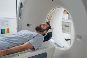
If you have never experienced an MRI before, then you might not be sure about what to expect. An MRI is a painless scan, though it can be uncomfortable for some people. The MRI machine is a long, tube-like structure that you enter while lying on a table. You have to stay very still during an MRI in order for the images to come out correctly. The MRI machine also uses heavy magnets to create the images, and the sound of the magnets moving around can be very loud and jarring. People who struggle with claustrophobia may have difficulty in a closed MRI machine. Thankfully, there are also open MRI machines and alternatives to MRI scans for people who cannot manage being in enclosed spaces.
The average MRI scan takes approximately 45 minutes, though the length of time really depends on what area of the body is being scanned. Because MRI scans use giant magnets to create the images, you cannot wear metal or jewelry during the scan. Your doctor will prepare you ahead of time with a list of appropriate clothing items and a checklist of what not to wear. However, MRI scans are generally safer and less risky because they do not expose you to any type of radiation.
How an MRI Can Help Inform Treatment
An MRI will translate images of your musculoskeletal system with a high level of detail. The high resolution of the MRI images provides your chiropractor with an accurate picture of your injury or condition. Your chiropractor will use the results of an MRI to inform their approach to your treatment and care. Our chiropractors and doctors at AICA Orthopedics use MRI scans all the time to help inform personalized treatment plans. AICA Orthopedics in Atlanta has walk-in chiropractors and on-site diagnostic imaging to make your visit as easy as possible. Contact us or stop by the office today and see one of our walk-in chiropractors and find out how an MRI scan can help support your treatment and care.
